Wolfram.com
WolframAlpha.com
WolframCloud.com
Wolfram Language
Example Repository
Ready-to-use examples of Wolfram Language
Primary Navigation
Categories
Algebra
Astronomy
Audio Processing
Calculus
Cellular Automata
Chemistry
Complex Systems
Computer Science
Computer Vision
Control Systems
Creative Arts
Data Science
Engineering
Finance & Economics
Finite Element Method
Food & Nutrition
Geography
Geometry
Graphs & Networks
Image Processing
Life Sciences
Machine Learning
Mathematics
Optimization
Physics
Probability & Statistics
Puzzles and Recreation
Quantum Computation
Signal Processing
Social Sciences
System Modeling
Tabular Processing
Text & Language Processing
Time-Related Computation
Video Processing
Visualization & Graphics
Alphabetical List
Submit a New Example
Learn More about
Wolfram Language
Related Pages
Related Categories
Chemistry
Life Sciences
Bacteriophage Head-Tail Connector Protein
Visualize and analyze a bacteriophage protein structure
Example Notebook
Open in Cloud
Download Notebook
Install the
ProteinVisualization
paclet and load the package:
I
n
[
1
]
:
=
P
a
c
l
e
t
I
n
s
t
a
l
l
[
"
W
o
l
f
r
a
m
C
h
e
m
i
s
t
r
y
/
P
r
o
t
e
i
n
V
i
s
u
a
l
i
z
a
t
i
o
n
"
]
;
N
e
e
d
s
[
"
W
o
l
f
r
a
m
C
h
e
m
i
s
t
r
y
`
P
r
o
t
e
i
n
V
i
s
u
a
l
i
z
a
t
i
o
n
`
"
]
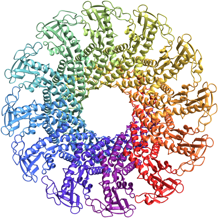
Visualize the structure of the
bacteriophage head-tail connector protein
as a ribbon diagram:
I
n
[
2
]
:
=
B
i
o
m
o
l
e
c
u
l
e
P
l
o
t
3
D
[
"
1
I
J
G
"
,
V
i
e
w
P
o
i
n
t
A
b
o
v
e
]
O
u
t
[
2
]
=
Generate a 3D plot of the backbone atoms and bonds of the protein, and color code the atoms by amino acid residues:
I
n
[
3
]
:
=
P
r
o
t
e
i
n
B
a
c
k
b
o
n
e
A
t
o
m
P
l
o
t
[
"
1
I
J
G
"
,
P
l
o
t
T
h
e
m
e
"
S
p
a
c
e
F
i
l
l
i
n
g
"
,
"
S
h
o
w
S
i
d
e
C
h
a
i
n
s
"
F
a
l
s
e
,
V
i
e
w
P
o
i
n
t
A
b
o
v
e
]
O
u
t
[
3
]
=
Show the
α
-carbon trace of the protein, color coded by chains:
I
n
[
4
]
:
=
A
l
p
h
a
C
a
r
b
o
n
P
a
t
h
P
l
o
t
3
D
[
"
1
I
J
G
"
,
"
B
a
c
k
b
o
n
e
C
o
l
o
r
R
u
l
e
s
"
{
"
C
h
a
i
n
s
"
,
A
u
t
o
m
a
t
i
c
}
,
V
i
e
w
P
o
i
n
t
A
b
o
v
e
]
O
u
t
[
4
]
=
To highlight the planar geometry of peptide bonds, show the amide planes along with the backbone atoms and bonds of the protein:
I
n
[
5
]
:
=
A
m
i
d
e
P
l
a
n
e
P
l
o
t
[
"
1
I
J
G
"
,
P
l
o
t
T
h
e
m
e
"
T
u
b
e
s
"
,
V
i
e
w
P
o
i
n
t
A
b
o
v
e
]
O
u
t
[
5
]
=
Highlight the planes that form
dihedral angles
ϕ
and
ψ
to get residue information and dihedral angle values upon mouse hover:
I
n
[
6
]
:
=
D
i
h
e
d
r
a
l
A
n
g
l
e
P
l
o
t
[
"
1
I
J
G
"
,
V
i
e
w
P
o
i
n
t
A
b
o
v
e
]
O
u
t
[
6
]
=
Plot the dihedral angles
ϕ
and
ψ
on a Ramachandran plot:
I
n
[
7
]
:
=
R
a
m
a
c
h
a
n
d
r
a
n
P
l
o
t
[
"
1
I
J
G
"
,
"
R
e
s
i
d
u
e
C
o
l
o
r
s
"
T
r
u
e
,
I
m
a
g
e
S
i
z
e
5
0
0
,
P
l
o
t
S
t
y
l
e
P
o
i
n
t
S
i
z
e
[
0
.
0
1
]
]
O
u
t
[
7
]
=
Create a
contact map
displaying residue-residue distances:
I
n
[
8
]
:
=
P
r
o
t
e
i
n
C
o
n
t
a
c
t
M
a
p
[
"
1
I
J
G
"
,
"
S
h
o
w
R
e
s
i
d
u
e
s
"
T
r
u
e
,
I
m
a
g
e
S
i
z
e
4
5
0
]
O
u
t
[
8
]
=
Visualize the residue-residue interactions within 8 Angstroms:
I
n
[
9
]
:
=
P
r
o
t
e
i
n
C
o
n
t
a
c
t
G
r
a
p
h
P
l
o
t
[
"
1
I
J
G
"
,
"
C
u
t
O
f
f
"
Q
u
a
n
t
i
t
y
[
8
,
"
A
n
g
s
t
r
o
m
s
"
]
,
"
S
c
a
l
e
d
V
e
r
t
e
x
S
i
z
e
"
T
r
u
e
,
"
E
d
g
e
C
o
l
o
r
F
u
n
c
t
i
o
n
"
T
r
u
e
,
I
m
a
g
e
S
i
z
e
5
0
0
,
V
e
r
t
e
x
S
i
z
e
3
0
]
O
u
t
[
9
]
=
External Links
Ramachandran Plot
1IJG
Source Metadata
Citation:
Simpson, Alan A., et al. "Structure determination of the head–tail connector of bacteriophage ϕ29." Acta Crystallographica Section D: Biological Crystallography 57.9 (2001): 1260-1269.
See Also
WolframChemistry/ProteinVisualization
Publisher Information
Contributed by:
Soutick Saha